Seed Implants (Brachytherapy)
Patient is placed under general anesthesia
A Catheter is placed through the urethra into the Bladder
Imaging is obtain using an Ultrasound placed in the patients rectum
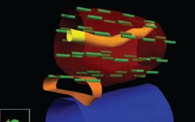
A 3 Dimensional representation of the Anatomy is generated in the treatment planning computer (Figure 4)
Multiple needle and seed approaches are evaluated and modeled in the computer before the optimal path and pattern is chosen
Thin empty needles are placed into the prostate
Radioactive seeds are placed individually into the prostate through the needles with a Mick Applicator
Doses are continually monitored and optimized throughout the procedure
At the completion of the procedure, all instruments are removed
The patients anesthesia is then reversed
Procedure
Fig 3. View of needle insertion into the prostate with Ultrasound probe in the rectum
SUMMARY
SIngle one hour Procedure
no incisions
go home the same day
source radioactivity is temporary

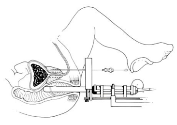
Fig 4. An example of a patient specific three dimensional prostate rendering (red), with rectum (blue), seminal vesicle (orange), individual radioactive seeds (green) and urethra (yellow)